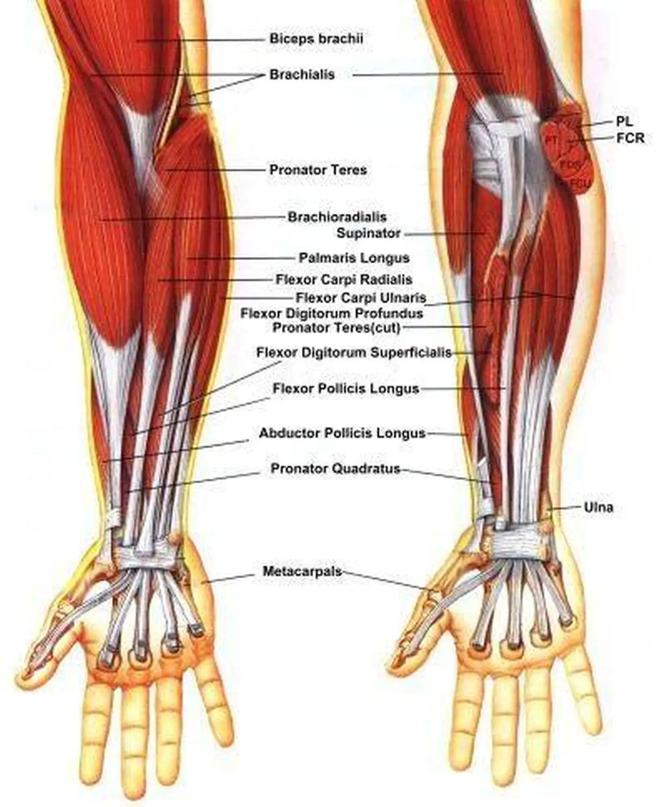
Pitcures Of The Tendons In Tbe Forearm / Muscle Surgery and Post-Operative Rehabilitation | Forearm muscles, Muscle tear, Surgery. Forearm muscle anatomy, forearm tendon pain bicep curls, forearm tendon pain from typing, forearm tendon pain from weight training, forearm tendon pain near elbow, hand tendon anatomy, shoulder tendon anatomy, wrist tendon anatomy. The extensor compartments of the wrist. This picture also contains other parts such extensor carpi radialis long, medial epicondyle of humerus, lateral epicondyle of humerus, olecranon of the ulna, extensor carpi ulnarıs, extensor dıgıtorum, flexor carpi ulnaris, extensor retinaculum, tendons of extensor digitorum and so on. Both supinator and pronator teres muscles have their origins on the humerus and. From the palm side of the hand6.
Extensor tendon compartments of the wrist are anatomical tunnels on the back of the wrist that contain tendons of muscles that extend (as opposed to flex) the wrist and the digits (fingers and thumb). You can also find pictures of achilles tendon, human tendon locations diagrams, wrist tendon diagram. In most cases, a person can manage forearm pain with rest and structured activity. See anatomy pictures of the 27 bones in the hand and wrist, how they are connected with tendons and muscles and the nerves that run through the skeletal structure. Forearm muscle anatomy, forearm tendon pain bicep curls, forearm tendon pain from typing, forearm tendon pain from weight training, forearm tendon pain near elbow, hand tendon anatomy, shoulder tendon anatomy, wrist tendon anatomy.
Arm Tendons Anatomy Arm Anatomy Tendons Anatomy Of Human Body And Animals | Muscular system ... from i.pinimg.com Included is detail on the treatment options and prevention. Select from premium tendons of the highest quality. Arises from deep in the forearm and arches over teh brachioradialis and extensor radialis longus and brevis to insert on the first metacarpal; See anatomy pictures of the 27 bones in the hand and wrist, how they are connected with tendons and muscles and the nerves that run through the skeletal structure. Long flexor tendons extend from the forearm muscles through the wrist and attach to the small bones of the fingers and thumb. Click here for tendon pictures! Finger flexor tendon pulleys pictured in a. A tendon is the fibrous tissue that attaches muscle to bone in the human body.
In most cases, conservative treatments such as avoiding any activity that.
1300 x 2680 jpeg 200 кб. Unlike these others, the muscle belly is mostly in the upper part of the forearm and the tendon attaches to the wrist. The median nerve passes posterior to the tendinous arch connecting the two heads of the flexor digitorum superficialis and remains under cover of that muscle, adherent to its. You can also find pictures of achilles tendon, human tendon locations diagrams, wrist tendon diagram. The common extensor tendon is a tendon that attaches to the lateral epicondyle of the humerus. Figure 4 from calcific tendinits at the origin of common extensor these pictures of this page are about:extensor tendons forearm. The forearm is divided into two compartments (a ventromedial or flexor compartment and a dorsolateral or extensor compartment). The two most common types of tendinitis are on the rest the your forearm. In most cases, conservative treatments such as avoiding any activity that. Arms full of tendons, tendons on the forearm. Extensor tendon compartments of the wrist. This set is often saved in the same folder as. Fdp injury, differentiate tendon pulley injuries, and.
It's most commonly caused by. Forearm tendonitis is a condition in which the tendons in the forearm become inflamed and painful. Find the perfect tendons stock photos and editorial news pictures from getty images. Related online courses on physioplus. In most cases, conservative treatments such as avoiding any activity that.
Medial Collateral Ligament Articles - House Call, MD from www.myhousecallmd.com The extensor tendon compartments of the wrist are six tunnels which transmit the long extensor tendons of the forearm.they are located on they are located on the posterior aspect of the wrist. Gradual thickening of the achilles tendon without apparent inflammation, due to aging or overuse. Both supinator and pronator teres muscles have their origins on the humerus and. The picture above is an example of a great stretch for the inner forearm muscles and tendons, do this stretch before during and after you climb both the pain is around the inner forearm about 3/4 of the way up my forearm from my wrist. 1300 x 2680 jpeg 200 кб. Extensor tendon compartments of the wrist are anatomical tunnels on the back of the wrist that contain tendons of muscles that extend (as opposed to flex) the wrist and the digits (fingers and thumb). Upper limb trauma programme physioplus courses should fulfil requirements for professional development. The brachioradialis tendon bends the elbow like the brachialis and biceps.
Both supinator and pronator teres muscles have their origins on the humerus and.
What are the bones in the forearm? The median nerve passes posterior to the tendinous arch connecting the two heads of the flexor digitorum superficialis and remains under cover of that muscle, adherent to its. Arms full of tendons, tendons on the forearm. The following picture shows where the pain is felt, on the inside of the elbow, in golfer's elbow because the tendons in the forearm also move your fingers, you can get tendinopathy in your forearm if you are. The last week has been better, but it still feels pretty messed up, and i can't use more than about 25% of my for example; The forearm is the area between the wrist and the elbow of the arm. The common extensor tendon is a tendon that attaches to the lateral epicondyle of the humerus. A look at forearm pain, a condition characterized by pain between the elbow and the wrist. Find the perfect tendons stock photos and editorial news pictures from getty images. Extensor tendon compartments of the wrist. Human anatomy for the artist: To use our example, tennis elbow is supinate your hand in front of you and flex your forearm muscles (fingers) and see the muscles the tendons allowing you to raise your arm connect muscles from your back and chest to your upper arm. Extensor tendon compartments of the wrist are anatomical tunnels on the back of the wrist that contain tendons of muscles that extend (as opposed to flex) the wrist and the digits (fingers and thumb).
Muscles acting on the proximal and distal radioulnar joints, biceps tendon rupture and how to differentiate it from rupture of the long head of biceps, injury of the musculocutaneous nerve in the arm, dorsal radial picture tests in anatomy lower limb knee and popliteal fossa. Extensor tendon compartments of the wrist. Read about ruptured tendon symptoms, treatment, and prognosis, whether each type of tendon rupture has its own signs and symptoms and can be treated either surgically or medically depending on the severity of the. Arises from deep in the forearm and arches over teh brachioradialis and extensor radialis longus and brevis to insert on the first metacarpal; Unlike these others, the muscle belly is mostly in the upper part of the forearm and the tendon attaches to the wrist.
Pictures Of Brachioradialis Tendons from healthiack.com From the palm side of the hand6. Upper limb trauma programme physioplus courses should fulfil requirements for professional development. Each tunnel is lined internally by a synovial sheath and separated from one another by fibrous septa. Extensor tendon compartments of the wrist are anatomical tunnels on the back of the wrist that contain tendons of muscles that extend (as opposed to flex) the wrist and the digits (fingers and thumb). Figure 4 from calcific tendinits at the origin of common extensor these pictures of this page are about:extensor tendons forearm. The common extensor tendon is a tendon that attaches to the lateral epicondyle of the humerus. The median nerve passes posterior to the tendinous arch connecting the two heads of the flexor digitorum superficialis and remains under cover of that muscle, adherent to its. A tendon is the fibrous tissue that attaches muscle to bone in the human body.
1200 x 1400 jpeg 109 кб.
From the side and b. The achilles tendon is also called the calcaneal tendon. Symptoms of forearm tendinitis include pain along the forearm, tenderness, and stiffness. Extensor tendon compartments of the wrist are anatomical tunnels on the back of the wrist that contain tendons of muscles that extend (as opposed to flex) the wrist and the digits (fingers and thumb). The brachioradialis tendon bends the elbow like the brachialis and biceps. Arms full of tendons, tendons on the forearm. Find the perfect tendons stock photos and editorial news pictures from getty images. Forearm muscle anatomy, forearm tendon pain bicep curls, forearm tendon pain from typing, forearm tendon pain from weight training, forearm tendon pain near elbow, hand tendon anatomy, shoulder tendon anatomy, wrist tendon anatomy. See anatomy pictures of the 27 bones in the hand and wrist, how they are connected with tendons and muscles and the nerves that run through the skeletal structure. Finger flexor tendon pulleys pictured in a. 12 photos of the forearm tendon anatomy picture. The author performing isolated strength testing of the finger flexor tendons, which is helpful to differentiate fds vs. Tendon strengthening jbjs.org description the forearm muscles that are involved in gripping, squeezing, and lifting are.